

Surgical Illustration:
Pediatric Right Ureteroureterostomy
Target audience
Surgery students
Supervisor
Professor Michael Corrin
Date completed
April 2020
Tools used
Pencil & Adobe Photoshop
Goal
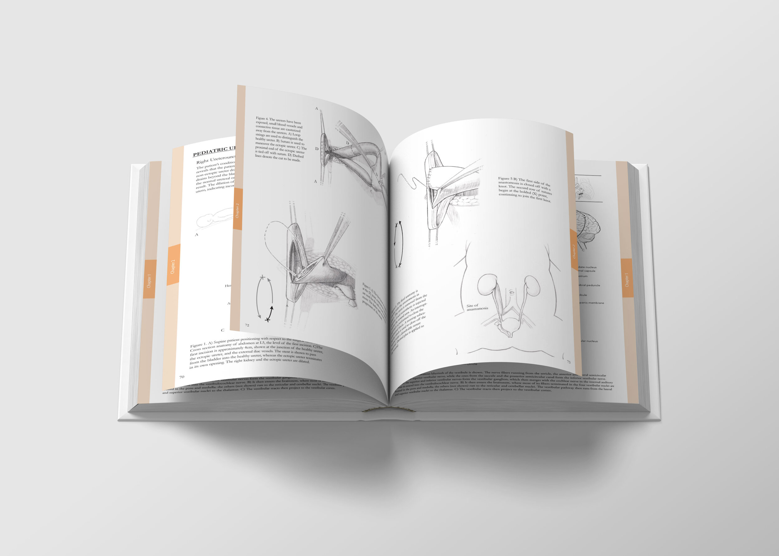
To illustrate the sequence of surgical steps involved in pediatric right ureteroureterostomy (retroperitoneal approach).


Process
Figure 1. Surgical Observation
We were granted the chance to enter the OR and observe the surgeries. The surgeons even answered our questions and offered explanations on the way. This was an incredible experience that allowed us to accumulate valuable surgical notes.
Figure 2. Cleaned up observation notes
The OR notes were cleaned up and the sketches were refined. The sequence of events was consolidated to keep the storytelling concise.

Figure 3. Establishing sketches
Each step was evaluated based on the level of acquired expertise of the audience. Common procedures (ex: incision method) were excluded to focus on ureteroureterostomy specific methodology.

Figure 5. Didactic Optimization
The orientation of the illustrations was changed from the ‘surgeon’s perspective’ to a traditional anatomical orientation to facilitate the learning process of surgical students.

Figure 6. Lighting and final details
A series of iterations were conducted to enhance the visual representation of tension (ureter being pulled) and interaction (the forceps pinching into the ureter).
Figure 7. Rendering
Pencil was used to mimic pen and ink hatching, due to the difficulty of acquiring materials during the onset of the pandemic.
Overall
Difficulties encountered:
Lack references for urology anatomy and surgery for infants.
Solution:
We gathered as many urology-related references as we could for adults and older children. We then made extrapolations based on that and the few infant urology anatomy references we had.
The surgeons were consulted at the end of the project to ensure the accuracy of the structures and procedures depicted.
References
Beattie, Edward, and Economou, Steven. An Atlas of Advanced Surgical Techniques. Philadelphia: W. B. Saunders Company, 1968.
Brooks, Shirley. Instrumentation for the Operating Room. Singapore: C. V. Mosby Company, 1989.
Burman, Curtis, and Kelly, Howard. Diseases of the Kidney, Ureters and Bladder: with Special Reference to the Disease in Women. New York: D. Appleton and Company, 1922.
Eycleshymer, Albert, and Schoemaker, Daniel. A Cross-Section Anatomy. New York: D. Appleton-Century Company, 1938.
Frick Hans, Krummer Benno, and Putz Reinhard. Wolf-Heidegger’s Atlas of Human Anatomy. Switzerland: S. KArger AG, 1990.
Rob, Charles, and Smith, Rodney. Operative Surgery: Urology. London: Butterworths, 1986.
Smith Joseph, Howards Stuart, Preminger Glenn, and Dmochowski Roger. Urology Surgery. Philadelphia: Elsevier, 2018.